Advanced diagnostic imaging is essential to guide therapy.
Today, cancer patients have more treatment options than ever before. The main purpose of imaging in oncology is early detection to enable interception if not prevention of full-blown disease, such as the appearance of metastases. Because biochemical changes occur well in advance of changes in anatomy, PET/CT molecular imaging provides especially early localization of disease. With this information, more optimal treatment selections can be made.
Errors and discrepancies in radiology practice are uncomfortably common [Insights Imaging. 2017 Feb; 8(1): 171–182]. Discordant interpretations in oncologist imaging is very high, ranging from 31 to 37%, and with expert second opinion interpretations resulting in a change in radiological staging in 19%, and change in patient treatment in up to 23%.
FDG, C11 - Acetate, Ga68-PSMA, F18 PyLarify PET/CT Imaging, Lutetium-177 (Lu177) PSMA Radioligand Therapy
Dr. Fabio Almeida is the only physician in the US who holds active dual board certifications in molecular imaging and integrative medicine. He is the former section chief of molecular imaging at the University of Arizona where he was also an associate professor of radiology and radiation oncology. As a recognized expert in PET/CT, he has been a leader in the development and interpretation of FDG, NaF18, C11-Acetate, Ga68 (Gallium-68) PSMA and F18 Pylarify PSMA PET/CT imaging studies, providing expert guidance on the selection of imaging studies, clinical trials and imaging over-reads. Advanced imaging navigation and interpretation is a key service of our Integrative Cancer Care. He is also one of the leading experts in radioligand therapy using Lu-177 PSMA, and coordinates this treatment for our clients for on-label and off-label use.


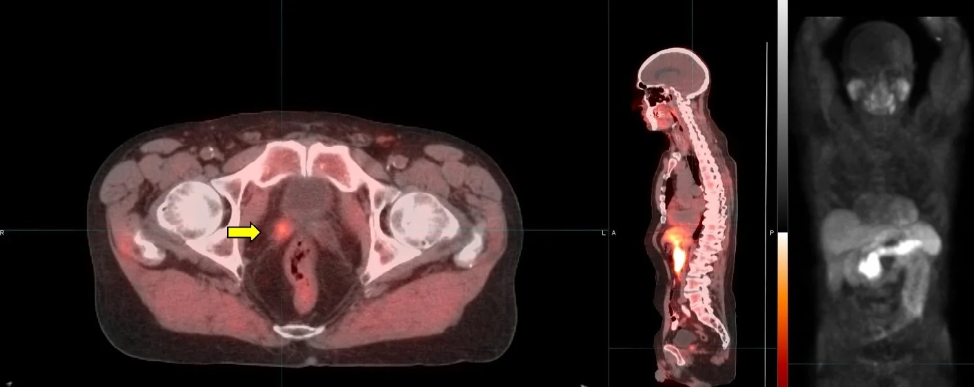
PET/CT Imaging - the basics
Positron Emission Tomography (PET/CT) is a major diagnostic imaging modality used predominantly in determining the presence and severity of cancers. It is currently the most effective way to check for cancer recurrence and to assess the response to treatment. Studies demonstrate that PET/CT offers significant advantages over other forms of imaging such as CT or MRI scans in diagnosing disease. Several hundred thousand clinical PET/CT patient studies are performed annually around the country.
PET/CT images demonstrate the chemistry of organs and other tissues such as tumors. A radiopharmaceutical, such as FDG (fluorodeoxyglucose), which includes both sugar (glucose) and a radionuclide (a radioactive element) that gives off signals, is injected into the patient and its emissions are measured by a PET/CT scanner.
While it varies based on the situation, there are several tumor types where PET/CT has proven to be critical:
Colorectal cancer
Melanoma
Head and neck cancer
Myeloma
Lung cancer
Breast cancer
Lymphoma
Prostate cancer (using PSMA agents)
PET/CT is considered particularly effective in identifying whether cancer is present or not, if it has spread, if it is responding to treatment, and if a person is cancer free after treatment. Cancers for which PET/CT is considered particularly effective include:
Early Detection: Because PET/CT images biochemical activity, it can accurately characterize a tumor as benign or malignant, thereby avoiding surgical biopsy when the PET/CT scan is negative. Conversely, because a PET/CT scan images the entire body, confirmation of distant metastasis can alter treatment plans in certain cases from surgical intervention to chemotherapy. Scientific studies estimate that PET/CT scans correctly identify lesions approximately 93% of the time. This accuracy in staging can also prevent patients from undergoing procedures that are not beneficial, helping them avoid unnecessary tissue or organ removal
Staging of Cancer: PET/CT is extremely sensitive in determining the full extent of disease, especially in lymphoma, malignant melanoma, breast, lung, colon and cervical cancers. Confirmation of metastatic disease allows the physician and patient to more accurately decide how to proceed with the patient’s management.
Checking for Recurrences: PET/CT is currently considered to be the most accurate diagnostic procedure to differentiate tumor recurrences from radiation necrosis or post-surgical changes. Such an approach allows for the development of a more rational treatment plan for the patient.
Assessing the Effectiveness of Chemotherapy or Immunotherapy : The level of tumor metabolism is compared on PET/CT scans taken before and after a chemotherapy cycle. A successful response seen on a PET/CT scan frequently precedes alterations in anatomy and would therefore be an earlier indicator of tumor response than that seen with other diagnostic modalities.